
Question asked: “…But how come there are electron microscope photos of them [viruses]?”
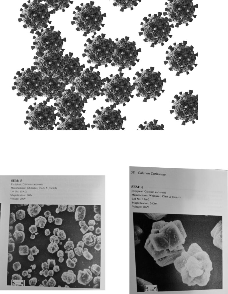
Bottom: Electron micrographs of calcium carbonate crystals/particles (Source: Handbook of Pharmaceutical Excipients, edited by A.H. Kibbe).
They aren’t. What is shown in those images is not an isolated virus but a mass of cell culture debris — gunk, frankly, the microscopic equivalent of a toilet flush. After the photo is taken, the debris is simply labeled “virus.” If it were truly a sample of an isolated virus, the image would show only viral particles, consistently and uniformly, just as electron micrographs of calcium carbonate show actual particles or crystals: every particle in the photo is calcium carbonate, taken from an isolated and purified sample.
People — including medical professionals — are fooled by “virus” images because they lack training in the science of isolation, preparation, and imaging. They do not understand how particles or their contents must be purified before they can be meaningfully photographed (even with an electron microscope). Too embarrassed to admit their ignorance, they instead are shouting louder and louder: “Here is the virus! Here is the virus!” But there is no virus. It is all fake and false.
The emperor has no clothes. All it will take is one honest observer to point out the obvious; then the crowd will see what was there (or not there) all along. Medical experts are not scientists — they are storytellers, and the story they have told is a tragic fiction.